by
John R. Fischer, Senior Reporter | August 22, 2023
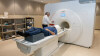
MIT researchers have developed a new technique combining physics and deep-learning for correcting motion in brain MR scans. (Photo courtesy of MIT)
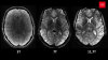
Patients undergoing MR scans must remain completely still in order to create quality, diagnostic images. With scans sometimes taking more than an hour, this is no small task, especially for children and patients with psychiatric and neurological conditions.
Utilizing deep learning with physics-based modeling, researchers at Massachusetts Institute of Technology (MIT) have developed a new approach for correcting motion to produce accurate brain scans. The scientists incorporated a deep network to reduce the joint search for image estimates and motion parameters to one for rigid motion parameters alone.
They compiled their findings in a paper, “Data Consistent Deep Rigid MRI Motion Correction.”


Ad Statistics
Times Displayed: 46612
Times Visited: 1410 MIT labs, experts in Multi-Vendor component level repair of: MRI Coils, RF amplifiers, Gradient Amplifiers Contrast Media Injectors. System repairs, sub-assembly repairs, component level repairs, refurbish/calibrate. info@mitlabsusa.com/+1 (305) 470-8013
“Not only is it excellent research work, but I believe these methods will be used in all kinds of clinical cases: children and older folks who can't sit still in the scanner, pathologies which induce motion, studies of moving tissue, even healthy patients will move in the magnet,” said Daniel Moyer, an assistant professor at Vanderbilt University in Nashville, where the paper was deemed the best oral presentation at the Medical Imaging with Deep Learning conference.
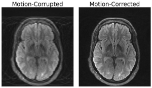
The network was trained on simulated, motion-corrupted k-space data indicative of typically-observed motions, and uses both of these factors as a basis for reconstructing images.
In addition to brain MR scans, the application could be used to reconstruct images distorted by motion in other body parts, including fetal MR, which often sees unpredictable motions that are challenging to account for when making diagnoses.
Prior research backs up the need for more accurate techniques to correct movement. In a 2022 study at Washington University School of Medicine in St. Louis, researchers developed a motion impact score that found movement deters the accuracy of functional MR brain scans, even after corrections are applied, according to a preprint published on bioRxiv.
In the last decade, several academic institutions and tech companies have developed products and techniques to address motion-related disturbances. In 2016, researchers at the University of Washington in Washington state created a functional MR technique that used multiple measurements to
correct motion and create 4D reconstructions of fetal brain activity.
Other approaches and solutions have instead focused on
preventing motion to begin with in the form of MR alert systems, or utilized AI to
improve image resolution to negate the effects of patients moving.
The study was supported by GE HealthCare and included computational hardware supplied by the Massachusetts Life Sciences Center.