by
Robin Lasky, Contributing Reporter | February 10, 2021
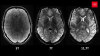
In a Phase III clinical trial, the use of MR for targeted biopsies of the prostate was found to be at least as effective in detecting cancerous growths as the standard ultrasound-guided transrectal approach, while offering a number of other advantages.
This study followed 453 patients of Canadian academic cancer centers who either had the benefit of MR prior to performing a biopsy or otherwise underwent the standard method of obtaining 12 prostate core samples aided by an ultrasound guided probe. Analysis of the results revealed that 5% more clinically significant prostate cancers were detected in the group that had the benefit of an MR prior to a targeted biopsy.
“My colleagues and I are thrilled about these results that show, without a doubt, that imaging and targeted biopsies are the future of prostate cancer diagnosis," said lead author, Dr. Laurence Klotz, in a statement. "We can catch more of the cancers we should be treating, avoid unnecessary treatment at the same time and improve the quality of life for our patients."


Ad Statistics
Times Displayed: 45747
Times Visited: 1372 MIT labs, experts in Multi-Vendor component level repair of: MRI Coils, RF amplifiers, Gradient Amplifiers Contrast Media Injectors. System repairs, sub-assembly repairs, component level repairs, refurbish/calibrate. info@mitlabsusa.com/+1 (305) 470-8013
A larger percentage of the MR group were determined to not require a biopsy, reducing clinically unnecessary biopsies of slowly progressing cancers by 55%. Further, those from the MR group that did undergo biopsies required significantly fewer samples taken, which reduces recovery time as well as the prevalence of common adverse events like hematuria and incontinence.
A biopsy performed with the use of an endorectal ultrasound-guided probe is currently the most common diagnostic method for identifying prostate cancer. The ultrasound probe is able to provide real time images of the prostate through the rectal wall, and similar to MR, can then be used to guide a needle insertion either through the rectum or the perineum in order to obtain a biopsy. However, compared with MR, ultrasound imaging is limited as to its utility in precisely identifying cancerous lesions.
In 2019, researchers from the U.K. and Canada conducted a
meta-analysis of seven prostate cancer studies, involving over 2,500 patients, which led them to conclude that a pre-biopsy MR led to less core samples being taken per procedure and thus, causing fewer adverse events.
The impact of adverse events associated with prostate cancer screening biopsies may significantly impact patient health outcomes. In 2017, a study was published of patients in Scotland analyzing the morbidity and
mortality risks posed by biopsies of the prostate. Its researchers found that following 120 days of a prostate biopsy procedure the risk of hospitalization and death, respectively, significantly increased regardless of whether the patient ended up with a cancer diagnosis or not.
The effectiveness of MR screening is also not without its own limitations and drawbacks. Diagnostic
results may significantly vary depending on the effectiveness of the urologist and technicians making use of it, and has been found to be unreliable when it comes to
assessing the size of cancerous growths. The technology is also significantly more expensive, compared with the standard ultrasound approach, contributing to reluctance by insurance companies to cover it, and as a result, fewer practitioners are experienced and trained in employing it effectively which may contribute to under-detection and treatment of clinically significant cancers.