by
Gus Iversen, Editor in Chief | December 23, 2020
From the November 2020 issue of HealthCare Business News magazine
NYU and Facebook make MR 4x faster without losing quality
Back in 2018, fastMRI, a collaboration between Facebook Artificial Intelligence Research and NYU Langone's Department of Radiology, released a large-scale, landmark MR data set with a plan to eventually power it with AI and make MR scans significantly faster.
In August they
published research in the
American Journal of Roentgenology showing how the team’s AI-generated MR scans are just as effective as a traditional scan, but with the added advantage of speed. Theoretically that means providers could scan more patients in a day and patients could experience less discomfort.


Ad Statistics
Times Displayed: 173735
Times Visited: 3176 For those who need to move fast and expand clinical capabilities -- and would love new equipment -- the uCT 550 Advance offers a new fully configured 80-slice CT in up to 2 weeks with routine maintenance and parts and Software Upgrades for Life™ included.
“This study is an important step toward clinical acceptance and utilization of AI-accelerated MR scans because it demonstrates for the first time that AI-generated images are essentially indistinguishable in appearance from standard clinical MR exams, and are interchangeable in regard to diagnostic accuracy,” said Michael P. Recht, professor of radiology at NYU Langone and lead study author. “This marks an exciting paradigm shift in how we are able to improve the patient experience and create images.”
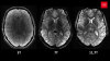
The open-source project comprised 1.5 million de-identified MR knee images from 10,000 scans, and raw measurement data from 1,600 scans, from which the researchers built a neural network and trained it. This included reconstruction of views missed from underlying image structure, similar to the way people interpolate sensory information.
In the study, musculoskeletal radiologists were asked to evaluate two sets of scan results per patient, including one from a standard MR exam and one from the fastMRI model, and were not told which one was created using AI. The images were examined for meniscal tears, ligament abnormalities, and cartilage defects, and also graded for quality. Both sets delivered the same results and led to the same diagnoses, but the radiologists said they preferred the AI image quality over the traditional images.