With DeHCA L&S physicians perform an initial optical examination with the objective to identify and locate areas of likely lesions, through the evaluation of the DeHCA biomarker. If any suspect areas are identified, physicians proceed to an ultrasound examination to exactly locate and qualify the lesions and eventually support a guided biopsy.
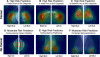
The optical information is acquired illuminating the breast with bottom-up 640nm red light, emitted by the LED’s positioned in the LED table (LED Plate). 640nm is the wavelength at which healthy and pathologic tissues show the maximum difference in transparency, which is lower in case of tumor neo-angiogenesis. Low transparency areas have presence of pathologic vessels.
The ultrasound information is used to exactly locate the lesion within the areas identified by the optical scan. The ultrasound morphological images and optical functional images can be directly compared.
HCB News: Can DeHCA provide a definitive diagnosis or would a patient with increased deoxyhemoglobin be referred for more follow up imaging? DG: With DeHCA L&S the diagnosis is made on the base of the ultrasound examination, while the optical examination is used as viewfinder. This procedure can be compared to what presently done with mammography and ultrasound. Therefore, we expect the DeHCA L&S diagnosis to be confirmed by biopsy, as in today’s clinical protocols.
HCB News: The system depends in part on a patented applicator that sort of vacuum seals onto the patients breast. What purpose does that serve and is it painful? DG: The evaluation of the concentration of deoxy-hemoglobin must be done when blood circulation in capillaries is temporarily stopped. This is achieved through a mild pressure exercised by the Applicator. The Applicator is a simple pressure/vacuum mechanism, at first glance similar to a bra, that makes the external ambient pressure to be superior to the pressure inside. In this way the Applicator membrane collapses on the breast and, perfectly adhering to it, provokes its immobilization on LED Plate and the temporary blockage of blood circulation in the neo-angiogenetic capillary vessels.
The mild applied pressure is about 20mmHg, absolutely painless, very much less than pressure applied to the breast with mammography.
HCB News: Is there any indication that breast density impacts the visualization of deoxyhemoglobin? DG: No. Breast density does not impact the visualization of deoxy-hemoglobin, because the 640nm red light is not stopped by glandular tissues.