Konica Minolta adds new features to SONIMAGE HSI Ultrasound with software upgrade
by
John R. Fischer, Senior Reporter | February 04, 2019
A new software upgrade has provided
new capabilities to the SONIMAGE HS1
Ultrasound System, including AI-assisted
voice control
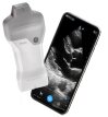
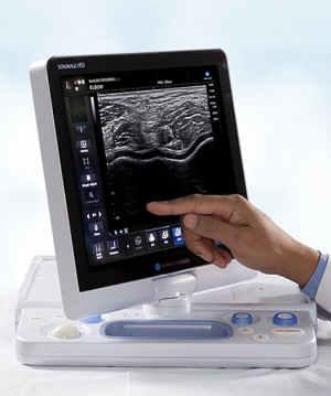
Users of Konica Minolta’s SONIMAGE HS1 Ultrasound System can expect greater functionality and new imaging capabilities for enhanced clinical workflow through the release of a new software upgrade.
Designed for musculoskeletal practitioners, the system now offers a variety of new features, including AI-assisted voice control functionality, that are meant to increase confidence in diagnoses and treatment management at the point-of-care.
“MSK practitioners expect their ultrasound systems to provide the best user experience, while raising the level of patient care,” Joan Toth, senior product marketing manager at Konica Minolta Healthcare Americas, told HCB News. “New system functionality, such as panoramic viewing and voice control, delivers improved workflow and enhanced image quality for diagnosis. This, in turn, provides a real-time, bedside, non-ionizing imaging modality for the patient and can often be used in place of more expensive imaging tests, such as MR.”
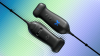
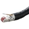
With AI-powered voice recognition technology, users can conduct hands-free operations during interventional procedures using simple voice commands to control system functions. A clinician, for instance, can hold the transducer in one hand and a needle or syringe in the other without the need for an assistant and maintaining the sterile field during exams.
The machine learning technology analyzes data from actual interactions to improve performance and accuracy, creating a voice control environment that supports proper operator ergonomics, and allows clinicians to feel more comfortable and focus more on the patient and procedure.
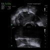
Users also have access to qualitative and quantitative imaging data through a new panoramic view that allows them to stitch together a series of images for a broader view of patient anatomy. With it, they can examine large-sized lesions, display an entire abnormality, build a cross-section image of a structure, and assess the relationship of two structures in a single image. This opens up the field of view for more accurate clinical diagnoses, measurements and interventions.
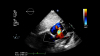
Another asset is the UltraAdjust one-touch image optimization feature, which can adjust depth to automatically alter multiple imaging parameters, such as frequency, focus and compounding, for greater image quality and resolution. This function, which can be activated with the new voice control operation as well, boosts confidence in diagnosis, therapeutic needle guidance and rehabilitation monitoring.
|
|
|
You Must Be Logged In To Post A Comment
|