Mount Sinai imaging symposium highlights latest research
by
Lisa Chamoff, Contributing Reporter | April 12, 2017
Multiparametric imaging of tumor habitats can help clinicians determine how best to treat patients and whether treatment is working.
Dr. Robert Gillies, chair of the Department of Cancer Imaging and Metabolism at the Moffitt Cancer Center, shared his research at the annual symposium hosted by the Translational and Molecular Imaging Institute (TMII) at the Icahn School of Medicine at Mount Sinai.
Clinicians at Moffitt don’t simply perform gene sequencing and select a drug therapy, but gather as much data from the patient as they can, Gillies told the audience via a web conference.
“We do pathomics on the histopathology, oftentimes with immunohistochemistry, we take a patient’s history into account, we do liquid biopsies if we can get them, we have lab data, molecular imaging and radiomic imaging, and we combine those using a mathematical model to predict where that tumor is, what it is and where it’s going,” Gillies said. “These models are actually nonlinear models, much like predicting hurricane paths. But the difference is [that] in predicting a hurricane path you’re just a passive participant, but in cancer we have the possibility of affecting the trajectory with therapy. We apply this therapy and most importantly we can collect these data here at the bottom, longitudinally to update the model to tell us whether or not we made the right call.”
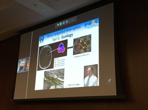
Gillies showed examples of how this process is being used to diagnose and treat myeloma and prostate cancer, at a time when targeted therapies are failing due to tumor evolution and ecology.
“The lack of a cure that we’re seeing with targeted therapies is due to the emergence of these preexisting resistant populations,” Gillies said. “Tumors have diverse habitats within ,and we can hope to see these and image these with multiparametric MRI.”
The seventh annual imaging symposium, attended by more than 200 faculty members and clinicians, was a way to create a forum to share research from invited speakers as well as the Mount Sinai community, and innovation in the different areas the institute covers, including cancer imaging, as well as neurology, cardiovascular disease and nanomedicine, or drug delivery, said Dr. Zahi Fayad, director of the TMII and a professor of medical imaging and bioengineering, radiology and cardiology at the Icahn School of Medicine.
After Gillies’s presentation, Dr. Paul Kennedy from the TMII shared research on new techniques to improve the diagnosis of liver cirrhosis, showing that 3-D magnetic resonance elastography (MRE) is a more effective modality than 2-D MRE.
Other invited speakers included Dr. Michael McConnell of Verily Life Sciences, who spoke about how the research organization is using technology to “re-engineer” health care, and Dr. Michael McMahon at Johns Hopkins School of Medicine, who discussed his work developing organic agents for Chemical Exchange Saturation Transfer (CEST) imaging.
During poster sessions, faculty members presented their work.
“It’s interactive,” Fayad said. “This is where people are building bridges, building collaborations. Maybe people come up with new ideas as we’re talking. That really fulfills our mission.”
|
|
|
You Must Be Logged In To Post A Comment
|