MR Spectroscopy Significantly Reduces Need for Breast Biopsy
by Barbara Kram, Editor | June 13, 2006
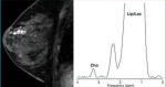
Suspicious lesion shown with
MR and magnified spectrum
MR and magnified spectrum
OAK BROOK, Ill.--In a study featured in the June issue of Radiology, researchers found that imaging suspicious breast lesions with magnetic resonance (MR) spectroscopy reduced the need for biopsy by 58 percent. The investigators, from Memorial Sloan-Kettering Cancer Center in New York, discovered that with the addition of MR spectroscopy to their breast MR imaging (MRI) protocol, 23 of 40 suspicious lesions could have been spared biopsy, and none of the resultant cancers would have been missed.
"All cancers in this study were identified with MR spectroscopy. There were no false-negative results," said Lia Bartella, M.D., lead investigator and assistant professor in the Department of Breast Imaging at Memorial Sloan-Kettering. "With the addition of MR spectroscopy to our breast MRI exam, we found that the number of biopsies recommended on the basis of MRI findings decreased significantly. These results should encourage more women to take this potentially life-saving test."
MRI is playing an increasingly important role in the screening of women at high risk for breast cancer. One drawback of the technology, however, has been a considerable number of breast biopsy procedures recommended on the basis of imaging findings, which turn out to be benign. With MR spectroscopy, the radiologist is able to see the chemical make-up of a tumor, so in most cases, he or she can tell without biopsy whether or not the lesion is cancerous.
"Breast tumors have elevated levels of choline compounds, which are a marker of an active tumor," Dr. Bartella said. "By performing a brief MR spectroscopy procedure after an MRI scan, which takes only 10 additional minutes, we can noninvasively see which tumors show elevated choline levels, and therefore which lesions are likely malignant. This eliminates the need for biopsy to find out what the tumor is made of."
In Dr. Bartella's study, 56 patients with 57 breast lesions underwent MRI first, followed by MR spectroscopy. Biopsy was performed after imaging, and results were compared.
At biopsy, there were 31 malignant lesions (54 percent) and 26 benign lesions (46 percent). All 31 malignant lesions were diagnosed correctly with MR spectroscopy (100 percent sensitivity), and 23 of 26 benign lesions were diagnosed correctly (88 percent specificity). The remaining three benign lesions showed elevated choline levels, even though they turned out to be benign at biopsy. Researchers are still exploring why certain types of benign lesions would have elevated choline levels, despite their non-malignant status.
1(current)