
The 10 biggest CT stories of 2020
December 16, 2020
MR might be involved in more cutting-edge research, but CT is the workhorse of advanced medical imaging. In 2020, the biggest stories had to do with utilization—the highs in 2019, a dramatic drop in 2020, access to screening, protocols for smoother operations, and an early role in diagnosing COVID-19.
Here, presented in semi-chronological order, are the 10 biggest CT stories of the year from our Daily News online.
 CT procedure volume in U.S. hit all-time high in 2019
While CT utilization plummeted in 2020, more than 90 million CT scans were performed throughout the U.S. in 2019, according to the 2019 CT Market Outlook Report, a study published in February 2020 by IMV Medical Information Division.
The estimated total of 91.4 million, an all-time record, represented a 3% increase from the 88.7 million recorded in 2018, and reflected volume increases indicated by respondents.
“The number of reported pelvis and abdomen procedures from 2011 to 2019 appears to have declined, but that may be due in part to the CMS policy, which bundled these procedures for reimbursement purposes,” Lorna Young, senior director of market research at IMV Medical Information Division, part of Science and Medicine Group, told HCB News. “Otherwise, we see procedure increases in most of the CT procedure types since 2011, including brain, CT Angiography (CTA), head and neck, spine, guided procedures, and low-dose CT used for lung screening.”
Among respondents, 79% of CT sites said their patient volume in 2019 for the modality was higher than their 2018 volume, a 12-point increase from the 67% in 2016 that indicated increases in patient volume.
The increase followed a slowdown in procedure volume between 2011 and 2018. Prior to that, CT procedures rose by 12% in average annual growth between 2001 and 2011, reaching 85.3 million in 2011. Growth declined from this peak by 5% to 80.6 million in 2012, with the study attributing the slowdown partially to changes in CMS reimbursement policy that involved bundling certain procedures together, such as abdomen and pelvis.
In addition, CMS further leveraged “Appropriate Use” criteria by mandating under the Protecting Access to Medicare Act of 2014 that physicians use a clinical decision support system to order imaging studies to minimize overutilization.
The rise in CT procedures was especially clear in emergency medicine, with about half of CT scans being performed on an emergency basis. An additional 30% were performed on outpatients, according to the study.
 Chest CT emerges as early COVID-19 diagnostic tool
In the early days of the COVID-19 pandemic, researchers from Wuhan, China published findings in Radiology showing that chest CT scans were more efficient than the lab tests available at the time for confirming a diagnosis.
Encompassing more than 1,000 patients, the assessment deemed CT to be the primary screening tool for COVID-19. At a time when therapeutic treatments and testing capabilities were in their infancy, imaging represented an invaluable tool for identifying and isolating infected patients from the healthy population.
The Chinese government began recognizing CT diagnoses for the coronavirus in February, having initially only recognized those confirmed by reverse-transcription polymerase chain reaction (RT-PCR) or gene sequencing for respiratory or blood specimens. Such tests were limited in sample collection, transportation and kit performance, with the total positive rate of RT-PCR for throat swab samples about 30%-60% at initial presentation. The low sensitivity of RT-PCR meant that a large number of COVID-19 patients would not be identified quickly and receive appropriate treatment, creating the risk of the virus spreading.
"Early diagnosis of COVID-19 is crucial for disease treatment and control,” wrote the researchers. “Compared to RT-PCR, chest CT imaging may be a more reliable, practical and rapid method to diagnose and assess COVID-19, especially in the epidemic area."
Around that time, early in the outbreak, CT equipment manufacturers like Siemens, GE, Canon, United Imaging Healthcare and Philips, donated millions of dollars in CT scanners and other equipment to support standard and makeshift hospitals diagnose patients quickly.
Meanwhile, Chinese and American researchers were collaborating to create a special report to educate radiologists on what signs to look for on CT scans when examining patients suspected of carrying the disease.
As time went on, the use of CT for diagnosing COVID-19 largely fell out of favor, as less costly imaging techniques and testing alternatives were developed.
Scan misinterpretation biggest cause of patient injury in diagnostic radiology
Evaluating closed malpractice claims in both diagnostic and interventional radiology, medical malpractice insurer The Doctors Company announced last December that most injuries took place in exams where misinterpretations occurred, which took place in 78 percent of cases — especially ones involving CT.
“Since the CT scan has become so ubiquitous and widely available, it has evolved into an essential tool in many imaging-based diagnoses,” Dr. Bradley Delman, vice chair for quality in radiology in the Mount Sinai Health System told HCB News. “Unlike the two-dimensional X-ray, a CT scan provides three-dimensional perspective, and with recent advances in resolution and imaging quality, a single CT exam often contains many hundreds of images. A subtle finding among such a large data set may be harder to detect overall. In addition, unlike the MR that may be used to refine a specific diagnosis, CT has become much more of a screening tool than it had been in the past.”
Delman reviewed the study for The Doctors Company along with other physician experts to form an accurate and unbiased understanding of what led to patient injuries. The most common type of misinterpretation was undiagnosed malignancy. CT scans were performed in 34 percent of the 78 percent of cases where injury was caused by scan misinterpretation.
For interventional radiology, technical performance was responsible for patient injuries in 76 percent of cases, most of which involved patients experiencing poor outcomes following invasive procedures. Technical performance led to negative results in 65 percent of cases where the correct procedure was performed appropriately, while only 11 percent of claims were due to poor technique or incorrect body site.
Darrell Ranum, vice president of the department of patient safety and risk management at The Doctors Company, chalks injuries in cases where procedures were appropriately executed up to risks of the operation. The findings, he told HCB News, highlight the importance of communication between radiologists and clinicians, as well as with patients prior to surgery and other procedures.
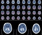 RSNA and ASNR create largest collection of annotated brain hemorrhage CT images to advance AI
In April, the RSNA and the American Society of Neuroradiology (ASNR), along with more than 60 volunteer neuroradiologists, unveiled what they’re calling the largest public collection of expert-annotated brain hemorrhage CT scans.
The collection, intended to speed up and support the creation of machine learning algorithms for diagnosing and characterizing brain hemorrhage, is a product of the latest edition of the RSNA Artificial Intelligence Challenge, in which participants were tasked with creating an algorithm to assist in identifying and characterizing intracranial hemorrhages from brain CT scans. The development process and observations of the data set were recorded in a paper published in Radiology: Artificial Intelligence.
“The value of this challenge is to create a data set that might lead to a generalizable solution, and the best way to do that is to train a model from data originating from multiple institutions that use a variety of CT scanners from various manufacturers, scanning protocols and a heterogeneous patient population,” said the paper’s lead author, Dr. Adam Flanders, a neuroradiologist and professor at Thomas Jefferson University Hospital, in a statement.
The release of the data set led to more than 22,000 submissions from 1,787 individual competitors in 1,345 teams from 75 countries in the challenge. The organizers expect its development by RSNA and a subspecialty like ASNR will help foster future collaborations.
“I was really impressed by the huge volunteer effort and the tremendous worldwide interest in this project,” Flanders said. “The data set we created for this challenge will endure as a valuable machine learning research resource for years to come.”
The data set will be used again in this year’s competition, which will be a collaboration between RSNA and the Society of Thoracic Radiology to detect and characterize pulmonary embolisms on chest CTs.
 USPSTF issues draft proposal expanding lung cancer screening access
The U.S. Preventive Services Task Force (USPSTF) expanded the eligibility criteria for low-dose CT lung cancer screenings in July with a draft recommendation lowering the age for testing people at high risk due to their smoking history from 55 to 50. It also recommended reducing the pack-years of smoking history that make an individual eligible for screening from 30 pack years to 20.
“Earlier detection of lung cancer ... that is the goal,” USPSTF member Dr. John B. Wong told HCB News. “The ability to detect lung cancer in earlier stages, when it’s more curable.”
More than 200,000 are diagnosed with lung cancer each year. Existing guidelines recommend that screening begin at 55 for those with 30 pack-years of smoking history. A pack year is a calculation of how much a person smokes, with one pack-year equivalent to smoking an average of 20 cigarettes, or one pack, per day for a year.
The draft is a Grade B recommendation but is based on evidence from Europe’s NELSON trial, the second largest randomized controlled trial to demonstrate a reduction in lung cancer mortality with CT screening of people at high risk, and the National Lung Cancer Screening Trial, a randomized U.S. trial conducted to determine whether screening with low-dose CT could reduce mortality from lung cancer.
The NLST trial, sponsored by the National Cancer Institute, found a 20% reduction in lung cancer mortality for annual screening over three years with low-dose CT scanning compared to chest radiography. The NELSON trial reaffirmed this finding, determining that low-dose CT screenings reduced lung cancer mortality by 24% in men and 33% in women compared to no screening. A follow-up NLST trial, also reaffirmed the results of both trials, finding a 26% reduction in lung cancer mortality in men and a 39% reduction in women.
The changes, according to Wong, would be especially helpful to African-Americans and women, who despite smoking fewer cigarettes than white men, are at higher risk of developing and dying from lung cancer.
Emory Healthcare shares tips for keeping CT exam schedules on time
Emory Healthcare in Atlanta, Georgia found, through a three-month retrospective analysis, that only 11% of its first-of-the-day CT exams were beginning on time, leading to unnecessary delays throughout the rest of the day. In July, they shared insight on their journey toward, and success with, resolving the problem.
The problem started at the very start of each day, with significant delays frequently impacting the first scheduled 8:30am outpatient case. From there, several downstream consequences would develop, including a snowball effect with delays in subsequent scheduled procedures resulting in an average delay of 71 minutes.
To address the issue, they turned to the lean and Six Sigma approach, a team method for improving performance, with the goal of standardizing their process and increasing on-time starts to more than 50% within 20 weeks.
Forming a team of proceduralists, technologists, nurses and administrators — with an administrator who had Six Sigma training leading the effort — Emory broke down and mapped out the entire process for admitting a patient into the CT room and beginning the procedure.
The team then performed a root cause analysis (RCA) of the various steps in the process. Most variability and lengthy times were found in nursing preparation of patients and coordination between nurses and CT technologists of when to let the patient enter the CT suite.
Emory orchestrated 14 different “tests of change” to address these issues, including equipping all nurses with access to the point-of-care INR testing device. Three changes received the most positive feedback:
1. Instituting daily morning huddles at 8:10am, with representatives from each procedural staff role.
2. Eliminating phone calls between nurses and CT techs used to assess readiness for accepting the patient.
3. Setting arrival time to 90 minutes prior to that first 8:30 a.m. appointment instead of an hour beforehand.
With the changes, mean turnaround time dropped from 71.5 minutes to 15.9, a gain of close to 78%. Exams starting on time — defined as within 15 minutes of an appointment — went from 11% to 82%, excluding outliers such as late arrivals.
CT X-ray cutting technique retains image quality at reduced dose
Researchers in London developed a technique for cutting a full X-ray beam during CT scans into thin beamlets, with the aim of reducing radiation exposure to the patient while retaining image quality.
"This new method fixes two problems,” said professor Sandro Olivo of University College London Medical Physics & Biomedical Engineering, senior author of a study on the technique, in a statement. “It can be used to reduce the dose, but if deployed at the same dose it can increase the resolution of the image.”
The method utilizes a mask with tiny slits that is placed over an X-ray beam. The slits allow for the beam to be divided into beamlets. Olivo and his colleagues tested their approach by imaging an object that was moved in a cycloidal motion to ensure quick and complete irradiation. The cycloidal method adds a simultaneous backward and forward motion to the rotation.
They then compared the outcomes to those of conventional CT scanning methods, in which a sample rotates as a full beam is directed on to it. Image quality was retained at a reduced dose, and image resolution was also found to be sharper, due to the part of the scanner that “reads” the information from the X-ray now being able to locate where the information came from more precisely. The sharpness can be easily adjusted using masks with different-sized apertures, freeing the resolution from the constraints of the scanner’s hardware.
The authors conducted the study with a micro CT scanner and believe it could potentially be adapted for medical scanners at some point in the future. Such an innovation, they say, could reduce the amount of radiation that millions of patients are exposed to, as about five million CT scans are performed annually in the U.K. and 80 million are performed in the U.S. CT scanning is believed to account for a quarter of Americans’ total exposure to radiation.
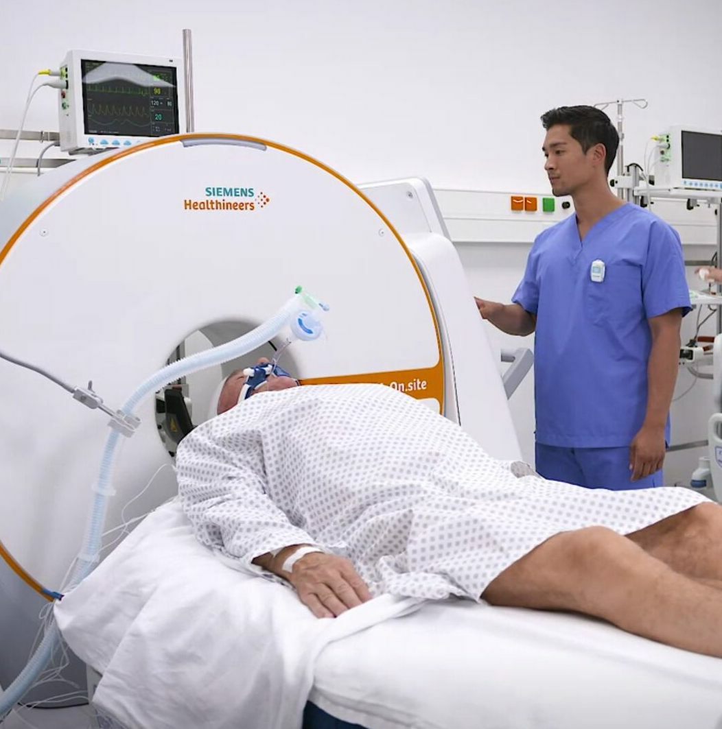
 FDA approves mobile head CT from Siemens
Siemens Healthineers announced FDA approval of a mobile CT scanner in early August that has the potential to reduce the risk of spreading COVID-19 in hospitals.
The 32-slice SOMATOM On.site mobile head CT scanner can image critically ill patients in the ICU without requiring them to leave their bed. Standard CT systems require staff to transport these patients to the radiology department, which poses significant risks.
“The minute that patient leaves their room, they are no longer in their isolation bubble,” Dena Cunningham, business development manager for mobile CT at Siemens, told HCB News. “Now they’re introduced to infection risk while they’re being transported.”
Sometimes up to five people, including a physician, are needed to transport these patients to the radiology department for their daily head scans. Aside from the infection risk, patient lines and/or ventilation tubes could be pulled out during transportation.
Cunningham noted the importance of also reducing infection risk among other patients frequenting the radiology department and staff. Keeping critically ill patients in the ICU could dramatically lower that risk.
The SOMATOM On.site first debuted at the 2019 RSNA annual meeting, where Cunningham said a lot of hospitals expressed interest in the system. In light of the pandemic, interest has increased.
“There are a lot of hospitals that are on budget freezes right now, but because this is considered such a unique product that can help take care of patients better by keeping them in their room, they’re talking about using emergency funding from the hospitals to purchase something like this,” she added.
 Radiologists make more errors when interpreting CT studies at night
In August, researchers at Mayo Clinic found that radiologists make more errors when interpreting CT studies overnight than they are when examining scans in the daytime.
The rate of errors was higher in off-hour body CT scans interpreted overnight, and particularly in the latter half of nighttime assignments. More broadly, the error rates were worse at night than during the day.
“The radiologists who were working overnight had schedules set up to give them ‘ample rest’," study author Dr. Maitray D. Patel, professor of radiology at Mayo Clinic Arizona, told HCB News. “They had 11 hours off before starting any assignment, and only worked a maximum of five nights in a row. The point is that someone has to read out the body CTs that occur after-hours, but are they susceptible to making more errors in doing that just because it is being done at night and they don’t always have a night schedule? Our study suggests that there is some impairment, even if they are well-rested for the night shift.”
Discrepancies affecting acute or follow-up clinical care were classified as errors. Daytime hours were between 7am and 5:59pm, while nighttime was between 6pm and 6:59am.
The team examined 10,090 body CT studies of the pelvis, abdomen or both, interpreted between July 2014 and June 2018. Scans were independently interpreted in-house and off-hours by radiologists who were part of a non-ACGME-accredited fellowship and were reviewed by an attending radiologist within 10 hours of initial interpretation, with the fellow interpretations submitted as complete final reports. Attending radiologists were specialists in body imaging, while the initial interpreting radiologists were fellows training in breast imaging (12 fellows), musculoskeletal imaging (eight fellows), or body MR (12 fellows).
Patel notes that body CT studies are notoriously one of the types of studies in which more errors are made by non-specialists, and says one possible reason may be the function of the number of different organs that must be evaluated.
CT scan volume falls nearly 3.7 million below expectations for 2020
In a year that was defined by COVID-19, it isn’t surprising that, from January through September, CT scans dropped by 19% compared to the previous year, according to analysis in the Journal of the American College of Radiology.
States on average reached a bottoming out point about 32 days into their emergency declaration and 12 days after stay-at-home orders were implemented. The average low point at that stage was a roughly 53% decline in CT volume.
A decline of 38,000 CT scans was seen on an average day during the worst parts of the crisis, with the largest drops seen in areas with higher dense populations and greater unemployment rates. CT visits were at about 84% of expected volume by September 30, indicating a “blunted recovery,” and that since CT volume never recovered to the levels predicted for it, a substantial proportion of care was not just deferred but not provided at all, according to lead author Dr. Matthew Davenport.
"It is likely that delayed and deferred care has had negative health implications for disease states unrelated to COVID-19," Davenport, who is service chief of Radiology and associate chair of operations at Michigan Medicine, told HCB News. "We do not yet have a complete picture of what those negative effects are, and we don't know whether those negative effects were outweighed by the positive effects of avoiding disease spread in the early months of the pandemic. Examples include delayed diagnosis and treatment of cancer."
The group analyzed data from 2,398 hospitals, academic facilities and freestanding imaging practices combined and scattered across all 50 states. They chalk the decline up to four factors: safety concerns, social distancing policies, staff reductions and the loss of health insurance by prospective patients.
Here, presented in semi-chronological order, are the 10 biggest CT stories of the year from our Daily News online.

While CT utilization plummeted in 2020, more than 90 million CT scans were performed throughout the U.S. in 2019, according to the 2019 CT Market Outlook Report, a study published in February 2020 by IMV Medical Information Division.
The estimated total of 91.4 million, an all-time record, represented a 3% increase from the 88.7 million recorded in 2018, and reflected volume increases indicated by respondents.
“The number of reported pelvis and abdomen procedures from 2011 to 2019 appears to have declined, but that may be due in part to the CMS policy, which bundled these procedures for reimbursement purposes,” Lorna Young, senior director of market research at IMV Medical Information Division, part of Science and Medicine Group, told HCB News. “Otherwise, we see procedure increases in most of the CT procedure types since 2011, including brain, CT Angiography (CTA), head and neck, spine, guided procedures, and low-dose CT used for lung screening.”
Among respondents, 79% of CT sites said their patient volume in 2019 for the modality was higher than their 2018 volume, a 12-point increase from the 67% in 2016 that indicated increases in patient volume.
The increase followed a slowdown in procedure volume between 2011 and 2018. Prior to that, CT procedures rose by 12% in average annual growth between 2001 and 2011, reaching 85.3 million in 2011. Growth declined from this peak by 5% to 80.6 million in 2012, with the study attributing the slowdown partially to changes in CMS reimbursement policy that involved bundling certain procedures together, such as abdomen and pelvis.
In addition, CMS further leveraged “Appropriate Use” criteria by mandating under the Protecting Access to Medicare Act of 2014 that physicians use a clinical decision support system to order imaging studies to minimize overutilization.
The rise in CT procedures was especially clear in emergency medicine, with about half of CT scans being performed on an emergency basis. An additional 30% were performed on outpatients, according to the study.

In the early days of the COVID-19 pandemic, researchers from Wuhan, China published findings in Radiology showing that chest CT scans were more efficient than the lab tests available at the time for confirming a diagnosis.
Encompassing more than 1,000 patients, the assessment deemed CT to be the primary screening tool for COVID-19. At a time when therapeutic treatments and testing capabilities were in their infancy, imaging represented an invaluable tool for identifying and isolating infected patients from the healthy population.
The Chinese government began recognizing CT diagnoses for the coronavirus in February, having initially only recognized those confirmed by reverse-transcription polymerase chain reaction (RT-PCR) or gene sequencing for respiratory or blood specimens. Such tests were limited in sample collection, transportation and kit performance, with the total positive rate of RT-PCR for throat swab samples about 30%-60% at initial presentation. The low sensitivity of RT-PCR meant that a large number of COVID-19 patients would not be identified quickly and receive appropriate treatment, creating the risk of the virus spreading.
"Early diagnosis of COVID-19 is crucial for disease treatment and control,” wrote the researchers. “Compared to RT-PCR, chest CT imaging may be a more reliable, practical and rapid method to diagnose and assess COVID-19, especially in the epidemic area."
Around that time, early in the outbreak, CT equipment manufacturers like Siemens, GE, Canon, United Imaging Healthcare and Philips, donated millions of dollars in CT scanners and other equipment to support standard and makeshift hospitals diagnose patients quickly.
Meanwhile, Chinese and American researchers were collaborating to create a special report to educate radiologists on what signs to look for on CT scans when examining patients suspected of carrying the disease.
As time went on, the use of CT for diagnosing COVID-19 largely fell out of favor, as less costly imaging techniques and testing alternatives were developed.
Scan misinterpretation biggest cause of patient injury in diagnostic radiology
Evaluating closed malpractice claims in both diagnostic and interventional radiology, medical malpractice insurer The Doctors Company announced last December that most injuries took place in exams where misinterpretations occurred, which took place in 78 percent of cases — especially ones involving CT.
“Since the CT scan has become so ubiquitous and widely available, it has evolved into an essential tool in many imaging-based diagnoses,” Dr. Bradley Delman, vice chair for quality in radiology in the Mount Sinai Health System told HCB News. “Unlike the two-dimensional X-ray, a CT scan provides three-dimensional perspective, and with recent advances in resolution and imaging quality, a single CT exam often contains many hundreds of images. A subtle finding among such a large data set may be harder to detect overall. In addition, unlike the MR that may be used to refine a specific diagnosis, CT has become much more of a screening tool than it had been in the past.”
Delman reviewed the study for The Doctors Company along with other physician experts to form an accurate and unbiased understanding of what led to patient injuries. The most common type of misinterpretation was undiagnosed malignancy. CT scans were performed in 34 percent of the 78 percent of cases where injury was caused by scan misinterpretation.
For interventional radiology, technical performance was responsible for patient injuries in 76 percent of cases, most of which involved patients experiencing poor outcomes following invasive procedures. Technical performance led to negative results in 65 percent of cases where the correct procedure was performed appropriately, while only 11 percent of claims were due to poor technique or incorrect body site.
Darrell Ranum, vice president of the department of patient safety and risk management at The Doctors Company, chalks injuries in cases where procedures were appropriately executed up to risks of the operation. The findings, he told HCB News, highlight the importance of communication between radiologists and clinicians, as well as with patients prior to surgery and other procedures.

In April, the RSNA and the American Society of Neuroradiology (ASNR), along with more than 60 volunteer neuroradiologists, unveiled what they’re calling the largest public collection of expert-annotated brain hemorrhage CT scans.
The collection, intended to speed up and support the creation of machine learning algorithms for diagnosing and characterizing brain hemorrhage, is a product of the latest edition of the RSNA Artificial Intelligence Challenge, in which participants were tasked with creating an algorithm to assist in identifying and characterizing intracranial hemorrhages from brain CT scans. The development process and observations of the data set were recorded in a paper published in Radiology: Artificial Intelligence.
“The value of this challenge is to create a data set that might lead to a generalizable solution, and the best way to do that is to train a model from data originating from multiple institutions that use a variety of CT scanners from various manufacturers, scanning protocols and a heterogeneous patient population,” said the paper’s lead author, Dr. Adam Flanders, a neuroradiologist and professor at Thomas Jefferson University Hospital, in a statement.
The release of the data set led to more than 22,000 submissions from 1,787 individual competitors in 1,345 teams from 75 countries in the challenge. The organizers expect its development by RSNA and a subspecialty like ASNR will help foster future collaborations.
“I was really impressed by the huge volunteer effort and the tremendous worldwide interest in this project,” Flanders said. “The data set we created for this challenge will endure as a valuable machine learning research resource for years to come.”
The data set will be used again in this year’s competition, which will be a collaboration between RSNA and the Society of Thoracic Radiology to detect and characterize pulmonary embolisms on chest CTs.

The U.S. Preventive Services Task Force (USPSTF) expanded the eligibility criteria for low-dose CT lung cancer screenings in July with a draft recommendation lowering the age for testing people at high risk due to their smoking history from 55 to 50. It also recommended reducing the pack-years of smoking history that make an individual eligible for screening from 30 pack years to 20.
“Earlier detection of lung cancer ... that is the goal,” USPSTF member Dr. John B. Wong told HCB News. “The ability to detect lung cancer in earlier stages, when it’s more curable.”
More than 200,000 are diagnosed with lung cancer each year. Existing guidelines recommend that screening begin at 55 for those with 30 pack-years of smoking history. A pack year is a calculation of how much a person smokes, with one pack-year equivalent to smoking an average of 20 cigarettes, or one pack, per day for a year.
The draft is a Grade B recommendation but is based on evidence from Europe’s NELSON trial, the second largest randomized controlled trial to demonstrate a reduction in lung cancer mortality with CT screening of people at high risk, and the National Lung Cancer Screening Trial, a randomized U.S. trial conducted to determine whether screening with low-dose CT could reduce mortality from lung cancer.
The NLST trial, sponsored by the National Cancer Institute, found a 20% reduction in lung cancer mortality for annual screening over three years with low-dose CT scanning compared to chest radiography. The NELSON trial reaffirmed this finding, determining that low-dose CT screenings reduced lung cancer mortality by 24% in men and 33% in women compared to no screening. A follow-up NLST trial, also reaffirmed the results of both trials, finding a 26% reduction in lung cancer mortality in men and a 39% reduction in women.
The changes, according to Wong, would be especially helpful to African-Americans and women, who despite smoking fewer cigarettes than white men, are at higher risk of developing and dying from lung cancer.
Emory Healthcare shares tips for keeping CT exam schedules on time
Emory Healthcare in Atlanta, Georgia found, through a three-month retrospective analysis, that only 11% of its first-of-the-day CT exams were beginning on time, leading to unnecessary delays throughout the rest of the day. In July, they shared insight on their journey toward, and success with, resolving the problem.
The problem started at the very start of each day, with significant delays frequently impacting the first scheduled 8:30am outpatient case. From there, several downstream consequences would develop, including a snowball effect with delays in subsequent scheduled procedures resulting in an average delay of 71 minutes.
To address the issue, they turned to the lean and Six Sigma approach, a team method for improving performance, with the goal of standardizing their process and increasing on-time starts to more than 50% within 20 weeks.
Forming a team of proceduralists, technologists, nurses and administrators — with an administrator who had Six Sigma training leading the effort — Emory broke down and mapped out the entire process for admitting a patient into the CT room and beginning the procedure.
The team then performed a root cause analysis (RCA) of the various steps in the process. Most variability and lengthy times were found in nursing preparation of patients and coordination between nurses and CT technologists of when to let the patient enter the CT suite.
Emory orchestrated 14 different “tests of change” to address these issues, including equipping all nurses with access to the point-of-care INR testing device. Three changes received the most positive feedback:
1. Instituting daily morning huddles at 8:10am, with representatives from each procedural staff role.
2. Eliminating phone calls between nurses and CT techs used to assess readiness for accepting the patient.
3. Setting arrival time to 90 minutes prior to that first 8:30 a.m. appointment instead of an hour beforehand.
With the changes, mean turnaround time dropped from 71.5 minutes to 15.9, a gain of close to 78%. Exams starting on time — defined as within 15 minutes of an appointment — went from 11% to 82%, excluding outliers such as late arrivals.
CT X-ray cutting technique retains image quality at reduced dose
Researchers in London developed a technique for cutting a full X-ray beam during CT scans into thin beamlets, with the aim of reducing radiation exposure to the patient while retaining image quality.
"This new method fixes two problems,” said professor Sandro Olivo of University College London Medical Physics & Biomedical Engineering, senior author of a study on the technique, in a statement. “It can be used to reduce the dose, but if deployed at the same dose it can increase the resolution of the image.”
The method utilizes a mask with tiny slits that is placed over an X-ray beam. The slits allow for the beam to be divided into beamlets. Olivo and his colleagues tested their approach by imaging an object that was moved in a cycloidal motion to ensure quick and complete irradiation. The cycloidal method adds a simultaneous backward and forward motion to the rotation.
They then compared the outcomes to those of conventional CT scanning methods, in which a sample rotates as a full beam is directed on to it. Image quality was retained at a reduced dose, and image resolution was also found to be sharper, due to the part of the scanner that “reads” the information from the X-ray now being able to locate where the information came from more precisely. The sharpness can be easily adjusted using masks with different-sized apertures, freeing the resolution from the constraints of the scanner’s hardware.
The authors conducted the study with a micro CT scanner and believe it could potentially be adapted for medical scanners at some point in the future. Such an innovation, they say, could reduce the amount of radiation that millions of patients are exposed to, as about five million CT scans are performed annually in the U.K. and 80 million are performed in the U.S. CT scanning is believed to account for a quarter of Americans’ total exposure to radiation.
Siemens Healthineers announced FDA approval of a mobile CT scanner in early August that has the potential to reduce the risk of spreading COVID-19 in hospitals.
The 32-slice SOMATOM On.site mobile head CT scanner can image critically ill patients in the ICU without requiring them to leave their bed. Standard CT systems require staff to transport these patients to the radiology department, which poses significant risks.
“The minute that patient leaves their room, they are no longer in their isolation bubble,” Dena Cunningham, business development manager for mobile CT at Siemens, told HCB News. “Now they’re introduced to infection risk while they’re being transported.”
Sometimes up to five people, including a physician, are needed to transport these patients to the radiology department for their daily head scans. Aside from the infection risk, patient lines and/or ventilation tubes could be pulled out during transportation.
Cunningham noted the importance of also reducing infection risk among other patients frequenting the radiology department and staff. Keeping critically ill patients in the ICU could dramatically lower that risk.
The SOMATOM On.site first debuted at the 2019 RSNA annual meeting, where Cunningham said a lot of hospitals expressed interest in the system. In light of the pandemic, interest has increased.
“There are a lot of hospitals that are on budget freezes right now, but because this is considered such a unique product that can help take care of patients better by keeping them in their room, they’re talking about using emergency funding from the hospitals to purchase something like this,” she added.

In August, researchers at Mayo Clinic found that radiologists make more errors when interpreting CT studies overnight than they are when examining scans in the daytime.
The rate of errors was higher in off-hour body CT scans interpreted overnight, and particularly in the latter half of nighttime assignments. More broadly, the error rates were worse at night than during the day.
“The radiologists who were working overnight had schedules set up to give them ‘ample rest’," study author Dr. Maitray D. Patel, professor of radiology at Mayo Clinic Arizona, told HCB News. “They had 11 hours off before starting any assignment, and only worked a maximum of five nights in a row. The point is that someone has to read out the body CTs that occur after-hours, but are they susceptible to making more errors in doing that just because it is being done at night and they don’t always have a night schedule? Our study suggests that there is some impairment, even if they are well-rested for the night shift.”
Discrepancies affecting acute or follow-up clinical care were classified as errors. Daytime hours were between 7am and 5:59pm, while nighttime was between 6pm and 6:59am.
The team examined 10,090 body CT studies of the pelvis, abdomen or both, interpreted between July 2014 and June 2018. Scans were independently interpreted in-house and off-hours by radiologists who were part of a non-ACGME-accredited fellowship and were reviewed by an attending radiologist within 10 hours of initial interpretation, with the fellow interpretations submitted as complete final reports. Attending radiologists were specialists in body imaging, while the initial interpreting radiologists were fellows training in breast imaging (12 fellows), musculoskeletal imaging (eight fellows), or body MR (12 fellows).
Patel notes that body CT studies are notoriously one of the types of studies in which more errors are made by non-specialists, and says one possible reason may be the function of the number of different organs that must be evaluated.
CT scan volume falls nearly 3.7 million below expectations for 2020
In a year that was defined by COVID-19, it isn’t surprising that, from January through September, CT scans dropped by 19% compared to the previous year, according to analysis in the Journal of the American College of Radiology.
States on average reached a bottoming out point about 32 days into their emergency declaration and 12 days after stay-at-home orders were implemented. The average low point at that stage was a roughly 53% decline in CT volume.
A decline of 38,000 CT scans was seen on an average day during the worst parts of the crisis, with the largest drops seen in areas with higher dense populations and greater unemployment rates. CT visits were at about 84% of expected volume by September 30, indicating a “blunted recovery,” and that since CT volume never recovered to the levels predicted for it, a substantial proportion of care was not just deferred but not provided at all, according to lead author Dr. Matthew Davenport.
"It is likely that delayed and deferred care has had negative health implications for disease states unrelated to COVID-19," Davenport, who is service chief of Radiology and associate chair of operations at Michigan Medicine, told HCB News. "We do not yet have a complete picture of what those negative effects are, and we don't know whether those negative effects were outweighed by the positive effects of avoiding disease spread in the early months of the pandemic. Examples include delayed diagnosis and treatment of cancer."
The group analyzed data from 2,398 hospitals, academic facilities and freestanding imaging practices combined and scattered across all 50 states. They chalk the decline up to four factors: safety concerns, social distancing policies, staff reductions and the loss of health insurance by prospective patients.