
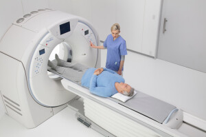
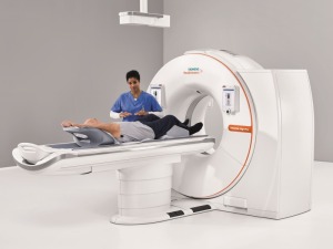
Canon Aquilion Precision
Spectral CT, workflow and dose reduction drive new CT scanner and software releases
October 15, 2018
by Lisa Chamoff, Contributing Reporter
Though the technology has been around for more than a decade, spectral CT is starting to gain ground in the CT market. Many of the new releases over the last year tout spectral imaging capabilities, meant for cancer detection and cardiovascular applications.
At the same time, manufacturers are improving workflow and software companies are helping providers get critical reimbursements while lowering radiation dose.
Here’s what’s new from the major CT players:
Canon Medical Systems USA
In April 2018, Canon Medical Systems USA received FDA clearance for its Aquilion Precision CT system. The scanner has an ultra-high-resolution detector with more than twice the resolution that is able to detect much smaller abnormalities – as small as 150 microns – and is part of a trend toward using CT for cancer detection and management, said Dominic Smith, senior director of the CT, MR and PET/CT business units for Canon Medical Systems USA Inc.
At last year’s RSNA, the company released the Aquilion Prime SP, a 160-slice all-purpose CT scanner that can be used for advanced cardiac care.
 Last fall the company also released an updated version of its Aquilion ONE GENESIS, with a technology that improves image quality in brain scans and for cardiac imaging.
Smith said the changes were prompted by new guidelines for the management of patients with stroke from the American Heart Association and American Stroke Association, which recommended CT over MR when triaging such cases.
The scanner’s Neuro FIRST MBIR application improves high-contrast spatial resolution and low contrast detectability in the brain, which allows physicians to possibly see early signs of stroke.
Aside from the buzz around the rise of spectral CT, a technology that has been around for the last decade, artificial intelligence is the next frontier for CT, and the company plans to release more information related to AI initiatives at this year’s RSNA.
“Our technology is built so that it’s AI ready,” Smith said. “AI is the next big trend that will impact all diagnostic imaging technology.”
CurveBeam
CurveBeam, which specializes in cone beam CT diagnostic imaging for the orthopedic market, received FDA clearance in May 2018 for its LineUP scanner, which provides weight-bearing imaging of the knee and lower extremities.
The scanners are specially designed for orthopedic practices, as they can plug into a regular wall outlet and don’t need a fully lead-lined room.
“Extremity scanners don’t require as much power as a full body CT,” said Vinti Singh, the marketing manager for CurveBeam. “It’s also convenient for patients, since they don’t have to travel to a separate imaging center or hospital for a scan.”
Some major medical centers have also purchased the scanner for their radiology departments because of the clinical advantage of standing exams.
“In a lot of lower extremity conditions, alignment is a very important aspect of understanding the condition,” Singh said.
 CurveBeam has also created a scanner for hand specialists, the InReach, which was FDA-cleared in May 2017. To use the scanner, the patient steps up to the scanner and places his or her hand inside a small gantry opening that is raised or lowered according to the patient's height.
CurveBeam’s products provide an advantage, as X-ray has limitations when imaging the extremities, Singh said.
“The foot and the hand are really complicated parts of the body,” Singh said. “It’s hard to see overlapping bone structures clearly on an X-ray. Incorrect tube head positioning can distort the anatomy. With CT, you have an exact three-dimensional view, with no distortions or overlaps.”
Singh said the next frontier for Curvebeam is a scanner that scans from the feet up to the hips, and the company’s engineers are already at work on a design.
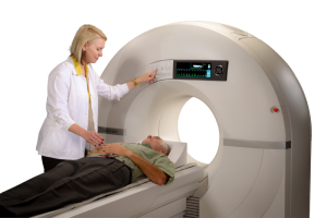
GE Healthcare
GE Healthcare’s latest CT release is the Revolution Frontier, a 128-slice scanner designed to provide spectral imaging at a more affordable price, according to the company.
The Frontier, FDA-cleared before last year’s RSNA, comes with a new tube and detector technology that improves overall image quality of spectral CT, said Scott Schubert, general manager of premium CT for GE Healthcare.
Peer reviewed papers looking at the Frontier technology show a 17 percent improvement in liver lesion characterization.
“That particular advantage means that 17 percent of your patients would not have to go to invasive follow-up testing,” Schubert said.
The company also doubled the overall workflow and speed of reconstruction, utilizing a partnership with AI computing company NVIDIA, allowing for the completion of post processing in five minutes, according to Schubert.
The company also recently released the CardioGraphe, a dedicated cardiovascular CT system. The scanner images the heart, coronary arteries and vascular structures and it can be used for structural heart procedures like transcatheter aortic valve replacement (TAVR).
“Facilities might not be able to afford top-of-the-line general purpose scanners,” Schubert said. “It is more of a middle price point with top-of-the-line performance for cardiovascular applications.”
The CardioGraphe is aimed at radiology practices with a high number of cardiac cases or imaging centers that want to set up chest pain management clinics, as well as for cardiology practices and in the cath lab.
“This is taking CT for the first time in a point-of-care setting, which is, of course, more comfortable for patients who have minor chest pain,” Schubert said. “Making the system more affordable in an outpatient setting is really the goal.”
 Since RSNA, GE has also released three new advanced clinical applications to improve diagnosis and accelerate workflow for CT.
The first, GSI Fat, is a fat quantification tool to determine whether a patient is susceptible to fatty liver disease, which is a precursor to liver cancer.
“Fat has been demonstrated to be very useful to show patients who may be pre-symptomatic,” Schubert said. “The gold standard has been MR in the past, but that is not for a broad population of patients.”
GE also released a physician visualization tool for 4D myocardial perfusion scans that Schubert said can “image the heart in one heartbeat, but also scan the heart over time and see perfusion of the myocardia.”
The third new application is for planning minimally-invasive mitral valve replacement procedures. The application is similar to GE’s aortic valve replacement planning tool, which evaluates the insertion point, makes sure the valve won’t dislodge any plaque and ensures that the size of the valve is adequate.
In the last year, GE also began offering what it calls Smart Subscription, which provides access in the cloud to the latest software updates for a facility’s entire fleet of CT scanners.
“This keeps all your scanners and software applications up to date with the latest upgrade subscription for the applications,” Schubert said.
Hitachi
At last year’s RSNA, Hitachi released a compact, economical CT scanner for the value-oriented market of community hospitals called the Supria True64.
The scanner is an upgrade from the Supria 16-slice model and it’s called the True64 because “it is a CT that really has 64 discrete detector channels,” said Mark Silverman, manager of CT marketing for Hitachi. “Being a true 64 gives it some speed of scanning and resolution advantages.”
 For the premium market, many manufacturers design a true 64-slice scanner, but for the economy market, they create a 32-slice CT that uses software upgrades to get the 64 slices, Silverman said.
“The 16-slice market is beginning to fade away,” Silverman said. “The true 64 is becoming the new 16.”
Supria True64 also comes with a new environmental efficiency feature called Eco-Mode that reduces power consumption by up to 55 percent when the scanner is idle.
MedicVision
Back in early 2015, the Centers for Medicare and Medicaid Services (CMS) announced reimbursement for low-dose CT lung cancer screenings for certain Medicare beneficiaries.
The screening presents a new potential source of income for imaging centers, but the scans cannot exceed radiation levels of 1.5 mSv in order to be eligible for reimbursement, which can be hard to achieve with older or lower-level CT scanners, said Eyal Aharon, the chief executive officer of MedicVision.
“In many places where they use older machines, they cannot get reimbursement,” Aharon said.
Last year, the company released SafeCT LS, a cloud-based solution – there is no hardware or on-site software installation – that reprocesses the low-dose scans to help facilities get under the 1.5 mSv threshold while retaining image quality. A Cloud-based solution eliminates the investment in equipment and facilities pay for SafeCT LS per scan.
While lung cancer screening programs have been slow to adoption, Aharon said the company has noticed an increase in the market in the last year or so, and thus the need for such a solution.
Neusoft
Around last year’s RSNA, Neusoft received FDA clearance for its NeuViz Prime 128-slice dual energy CT scanner with spectral imaging capabilities.
What makes the system unique is a newly designed X-ray tube that removes heat faster than it's introduced, said Keith Mildenberger, the CT product manager for Neusoft.
The NeuViz Prime offers a 0.259 second rotation speed, allowing for motion suppression that is ideal for pediatric imaging, as well as trauma and cardiac cases.
“In pediatrics, motion is the enemy,” Mildenberger said. “It will be a really great pediatric system and should have the potential to do very good cardiac imaging as well.”
The system also has the ability to upgrade to perform spectral imaging, which Mildenberger said has a big future in cardiac imaging and virtual colonoscopy.
 “The market we’re in is very dynamic and changing very rapidly,” Mildenberger said. “We have all the tools in place to support the clinical applications for spectral CT.”
PACSHealth LLC
In the last year, PACSHealth LLC, a radiology informatics software company, added new features to version 2.5.4 of its DoseMonitor software. The software can now perform organ dose modeling both pre- and post-scan, using phantom sets from National Cancer Institute.
In July, the company added Modality Utilization reporting, which monitors the time that the CT scanner is actually utilized based on scan time, not time the room is blocked. This information automatically gets sent to all customers for better planning of exams.
"Now we can retrospectively analyze how long the exam took versus what it was scheduled for," said Mike Battin, chief operating officer of PACSHealth.
PACSHealth also recently introduced its Global Dose Registry, a feature that is integrated into DoseMonitor and that allows clinicians to compare the radiation dose of an exam to a global database of millions of similar studies done at healthcare facilities around the world.
Battin said that this is a big improvement over existing data aggregation sources that require separate data collection and manual reporting.
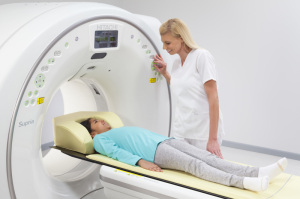
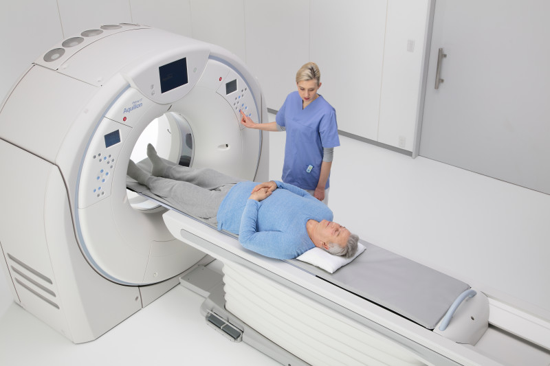
Philips Healthcare
At last year’s RSNA, Philips introduced its IQon Elite Spectral CT scanner, a premium product that is ideal for oncology applications, as well as for cardiology and trauma settings.
The scanner can detect different levels of tissue composition, distinguishing between healthy and unhealthy cells, allowing for doctors to be more confident in their diagnoses, said Karim Boussebaa, head of CT and AMI for Philips.
“The advantage is in the ability to look at tissue composition at various levels of chemistry,” Boussebaa said.
The scanner also provides faster reconstruction and image quality improvements, decreasing the need to reimage patients, according to Boussebaa.
 The IQon Elite is the world’s first and only spectral detector-based CT scanner, according to the company, and is part of a shift toward the technology.
“As price goes down, spectral will become new normal,” Boussebaa said.
Siemens Healthineers
It’s been a busy year for the Siemens Healthineers CT segment. In April 2018, the company received FDA clearance for the SOMATOM go.All and SOMATOM go.Top systems, the latest addition to its SOMATOM family of CT scanners.
The scanners include a redesigned tablet workflow that allows technologists to spend more time with patients, a development that came after the company engaged with more than 500 customers from around the world in co-creation sessions.
“We believe this is a major enhancement,” said Matthew Dedman, U.S. marketing director for CT for Siemens Healthineers North America.
Along with the tablet workflow comes intelligent automation, including the ability to set up the scan range and the reconstruction field of view automatically, for greater standardization and consistency of results, Dedman said.
The scanners also come with a larger 75-kilowatt generator, for busier and more complex environments.
“We enhanced the horsepower of those systems to enable more advanced exams, such as emergency department patients and cardiac,” Dedman said.
 Both systems utilize the new Athlon X-ray tube, which Dedman said offers its highest mA output of 825 mA at its lowest kV settings, allowing radiation dose to be tailored to each patient.
“The industry as a whole has done a good job in investing in iterative reconstruction,” Dedman said. “Additionally, we have invested heavily into hardware-based dose reduction technologies as well. If we can optimize our hardware, so that prior to any iterative reconstruction we are delivering the lowest dose, high-quality image, that means we’re less reliant on IR algorithms and can maintain a more natural image impression for the radiologist.”
In April, the company also received FDA clearance for its SOMATOM Edge Plus and SOMATOM Force CT systems. Both scanners feature Fully Assisting Scanner Technologies (FAST) Integrated Workflow with what the company calls the FAST 3D Camera, a patient positioning system that uses artificial intelligence.
Historically, patient positioning has been a manual task, with the height of the technologist impacting the patient position, Dedman explained.
“A six-four tech versus a five-six tech will have a different visual perception of what the ISO center is,” Dedman said.
The FAST 3D camera, mounted above the table, uses an AI algorithm to do correct positioning for the exam, reducing variability among exams.
“When we looked at areas to develop new technologies, we’re always looking for ways to improve standardization and increase consistency for our customers,” Dedman said.
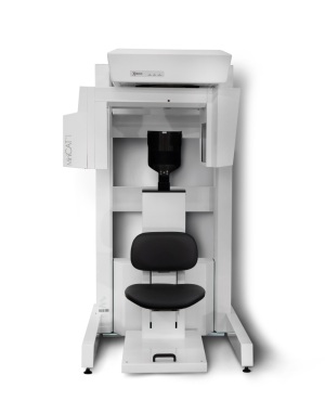 Xoran Technologies LLC
Xoran specializes in point-of-care CT scanners that are utilized in doctor's offices as well as the operating room. The company’s flagship product is the MiniCAT, which is primarily used in ENT and allergy offices for CT scans of the ears and sinuses.
In April 2018, the company released the MiniCAT 2020, a scanner that is further optimized for CT imaging within the workflow of the ENT clinic.
The scanner can differentiate between bone, liquid and air, giving clinicians the ability to detect disease, infection, obstruction and other ailments, said Russell Jahnke, director of product management for Xoran Technologies.
“We really looked at what patients are expecting,” Jahnke said. “When they go see their ENT, they really want to see things taken care of in as few visits as possible. The point-of-care imaging the MiniCAT offers in the ENT office is exactly that.”
In the middle of last year, the company introduced the xCAT, a portable head CT scanner for use in the OR, allowing surgeons access to CT imaging during surgery. This year, Xoran also released the xCAT IQ, a portable intraoperative CT designed for neurosurgeons to use in both the OR and the ICU.
“If a neurosurgery patient is in the ICU, the current standard-of-care is to pack them up and take them to the CT suite,” Jahnke said. “With the xCAT IQ, you can bring the device to the bedside.”
Earlier in 2018, Xoran debuted what it calls “Ace Mode” on its MiniCAT IQ sinus and temporal bone scanner used in the doctor's office. When MiniCAT is in ACE Mode, it uses patient-specific data obtained during the pre-scan setup to automatically present a patient profile that helps the clinician select the just-right patient CT scan.
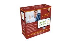 Zetta Medical Technologies
In July 2017, Zetta Medical Technologies, a third-party service provider that also develops CT software, released Z-DOSE RP, a dose reporting platform. The software connects with the ACR dose registry and also allows facilities to track dose data by study type over multiple scanners and locations.
“A lot of the legacy systems don’t have those capabilities,” said Mike Ghazal, the chief executive officer of Zetta Medical Technologies. “Even the new scanners will generate a structured report, but don’t generate a dose report for the director of radiology to view and track data. Ours is more of a dashboard comparison to analyze dose data over time using multiple criteria. In addition to CTDi and DLP values, it also includes comparisons by exam type, scanner type, referring physician, etc.”
The product is designed for large university hospitals and imaging centers, and is designed to work with the scanners from the major manufacturers, including Canon, GE, Philips and Siemens.
“Once they see the reports, they don’t want to go back to not using it,” Ghazal said. “They like the ability to see all this data and have it at their fingertips.”
The company has designed custom templates for its customers to allow them to view more data to their own specifications.
“Every place has its own set of protocols,” Ghazal said.
At the same time, manufacturers are improving workflow and software companies are helping providers get critical reimbursements while lowering radiation dose.
Here’s what’s new from the major CT players:
Canon Medical Systems USA
In April 2018, Canon Medical Systems USA received FDA clearance for its Aquilion Precision CT system. The scanner has an ultra-high-resolution detector with more than twice the resolution that is able to detect much smaller abnormalities – as small as 150 microns – and is part of a trend toward using CT for cancer detection and management, said Dominic Smith, senior director of the CT, MR and PET/CT business units for Canon Medical Systems USA Inc.
At last year’s RSNA, the company released the Aquilion Prime SP, a 160-slice all-purpose CT scanner that can be used for advanced cardiac care.
Canon Aquilion Prime SP
Smith said the changes were prompted by new guidelines for the management of patients with stroke from the American Heart Association and American Stroke Association, which recommended CT over MR when triaging such cases.
The scanner’s Neuro FIRST MBIR application improves high-contrast spatial resolution and low contrast detectability in the brain, which allows physicians to possibly see early signs of stroke.
Aside from the buzz around the rise of spectral CT, a technology that has been around for the last decade, artificial intelligence is the next frontier for CT, and the company plans to release more information related to AI initiatives at this year’s RSNA.
“Our technology is built so that it’s AI ready,” Smith said. “AI is the next big trend that will impact all diagnostic imaging technology.”
CurveBeam
CurveBeam, which specializes in cone beam CT diagnostic imaging for the orthopedic market, received FDA clearance in May 2018 for its LineUP scanner, which provides weight-bearing imaging of the knee and lower extremities.
The scanners are specially designed for orthopedic practices, as they can plug into a regular wall outlet and don’t need a fully lead-lined room.
“Extremity scanners don’t require as much power as a full body CT,” said Vinti Singh, the marketing manager for CurveBeam. “It’s also convenient for patients, since they don’t have to travel to a separate imaging center or hospital for a scan.”
Some major medical centers have also purchased the scanner for their radiology departments because of the clinical advantage of standing exams.
“In a lot of lower extremity conditions, alignment is a very important aspect of understanding the condition,” Singh said.

Curvebeam LineUP
CurveBeam’s products provide an advantage, as X-ray has limitations when imaging the extremities, Singh said.
“The foot and the hand are really complicated parts of the body,” Singh said. “It’s hard to see overlapping bone structures clearly on an X-ray. Incorrect tube head positioning can distort the anatomy. With CT, you have an exact three-dimensional view, with no distortions or overlaps.”
Singh said the next frontier for Curvebeam is a scanner that scans from the feet up to the hips, and the company’s engineers are already at work on a design.
GE Healthcare
GE Healthcare’s latest CT release is the Revolution Frontier, a 128-slice scanner designed to provide spectral imaging at a more affordable price, according to the company.
The Frontier, FDA-cleared before last year’s RSNA, comes with a new tube and detector technology that improves overall image quality of spectral CT, said Scott Schubert, general manager of premium CT for GE Healthcare.
Peer reviewed papers looking at the Frontier technology show a 17 percent improvement in liver lesion characterization.
“That particular advantage means that 17 percent of your patients would not have to go to invasive follow-up testing,” Schubert said.
The company also doubled the overall workflow and speed of reconstruction, utilizing a partnership with AI computing company NVIDIA, allowing for the completion of post processing in five minutes, according to Schubert.
The company also recently released the CardioGraphe, a dedicated cardiovascular CT system. The scanner images the heart, coronary arteries and vascular structures and it can be used for structural heart procedures like transcatheter aortic valve replacement (TAVR).
“Facilities might not be able to afford top-of-the-line general purpose scanners,” Schubert said. “It is more of a middle price point with top-of-the-line performance for cardiovascular applications.”
The CardioGraphe is aimed at radiology practices with a high number of cardiac cases or imaging centers that want to set up chest pain management clinics, as well as for cardiology practices and in the cath lab.
“This is taking CT for the first time in a point-of-care setting, which is, of course, more comfortable for patients who have minor chest pain,” Schubert said. “Making the system more affordable in an outpatient setting is really the goal.”

GE CardioGraphe
The first, GSI Fat, is a fat quantification tool to determine whether a patient is susceptible to fatty liver disease, which is a precursor to liver cancer.
“Fat has been demonstrated to be very useful to show patients who may be pre-symptomatic,” Schubert said. “The gold standard has been MR in the past, but that is not for a broad population of patients.”
GE also released a physician visualization tool for 4D myocardial perfusion scans that Schubert said can “image the heart in one heartbeat, but also scan the heart over time and see perfusion of the myocardia.”
The third new application is for planning minimally-invasive mitral valve replacement procedures. The application is similar to GE’s aortic valve replacement planning tool, which evaluates the insertion point, makes sure the valve won’t dislodge any plaque and ensures that the size of the valve is adequate.
In the last year, GE also began offering what it calls Smart Subscription, which provides access in the cloud to the latest software updates for a facility’s entire fleet of CT scanners.
“This keeps all your scanners and software applications up to date with the latest upgrade subscription for the applications,” Schubert said.
Hitachi
At last year’s RSNA, Hitachi released a compact, economical CT scanner for the value-oriented market of community hospitals called the Supria True64.
The scanner is an upgrade from the Supria 16-slice model and it’s called the True64 because “it is a CT that really has 64 discrete detector channels,” said Mark Silverman, manager of CT marketing for Hitachi. “Being a true 64 gives it some speed of scanning and resolution advantages.”
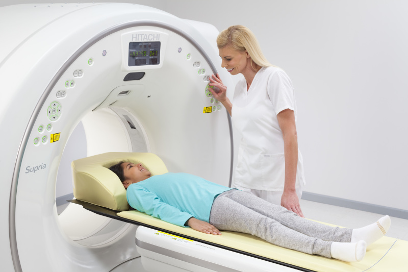
Hitachi Supria True64
“The 16-slice market is beginning to fade away,” Silverman said. “The true 64 is becoming the new 16.”
Supria True64 also comes with a new environmental efficiency feature called Eco-Mode that reduces power consumption by up to 55 percent when the scanner is idle.
MedicVision
Back in early 2015, the Centers for Medicare and Medicaid Services (CMS) announced reimbursement for low-dose CT lung cancer screenings for certain Medicare beneficiaries.
The screening presents a new potential source of income for imaging centers, but the scans cannot exceed radiation levels of 1.5 mSv in order to be eligible for reimbursement, which can be hard to achieve with older or lower-level CT scanners, said Eyal Aharon, the chief executive officer of MedicVision.
“In many places where they use older machines, they cannot get reimbursement,” Aharon said.
Last year, the company released SafeCT LS, a cloud-based solution – there is no hardware or on-site software installation – that reprocesses the low-dose scans to help facilities get under the 1.5 mSv threshold while retaining image quality. A Cloud-based solution eliminates the investment in equipment and facilities pay for SafeCT LS per scan.
While lung cancer screening programs have been slow to adoption, Aharon said the company has noticed an increase in the market in the last year or so, and thus the need for such a solution.
Neusoft
Around last year’s RSNA, Neusoft received FDA clearance for its NeuViz Prime 128-slice dual energy CT scanner with spectral imaging capabilities.
What makes the system unique is a newly designed X-ray tube that removes heat faster than it's introduced, said Keith Mildenberger, the CT product manager for Neusoft.
The NeuViz Prime offers a 0.259 second rotation speed, allowing for motion suppression that is ideal for pediatric imaging, as well as trauma and cardiac cases.
“In pediatrics, motion is the enemy,” Mildenberger said. “It will be a really great pediatric system and should have the potential to do very good cardiac imaging as well.”
The system also has the ability to upgrade to perform spectral imaging, which Mildenberger said has a big future in cardiac imaging and virtual colonoscopy.
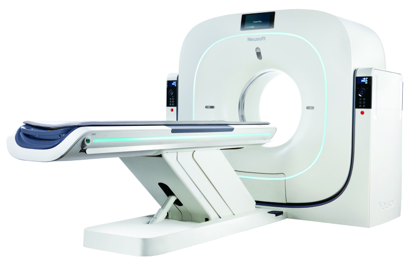
Neusoft NeuViz Prime
PACSHealth LLC
In the last year, PACSHealth LLC, a radiology informatics software company, added new features to version 2.5.4 of its DoseMonitor software. The software can now perform organ dose modeling both pre- and post-scan, using phantom sets from National Cancer Institute.
In July, the company added Modality Utilization reporting, which monitors the time that the CT scanner is actually utilized based on scan time, not time the room is blocked. This information automatically gets sent to all customers for better planning of exams.
"Now we can retrospectively analyze how long the exam took versus what it was scheduled for," said Mike Battin, chief operating officer of PACSHealth.
PACSHealth also recently introduced its Global Dose Registry, a feature that is integrated into DoseMonitor and that allows clinicians to compare the radiation dose of an exam to a global database of millions of similar studies done at healthcare facilities around the world.
Battin said that this is a big improvement over existing data aggregation sources that require separate data collection and manual reporting.
Philips Healthcare
At last year’s RSNA, Philips introduced its IQon Elite Spectral CT scanner, a premium product that is ideal for oncology applications, as well as for cardiology and trauma settings.
The scanner can detect different levels of tissue composition, distinguishing between healthy and unhealthy cells, allowing for doctors to be more confident in their diagnoses, said Karim Boussebaa, head of CT and AMI for Philips.
“The advantage is in the ability to look at tissue composition at various levels of chemistry,” Boussebaa said.
The scanner also provides faster reconstruction and image quality improvements, decreasing the need to reimage patients, according to Boussebaa.
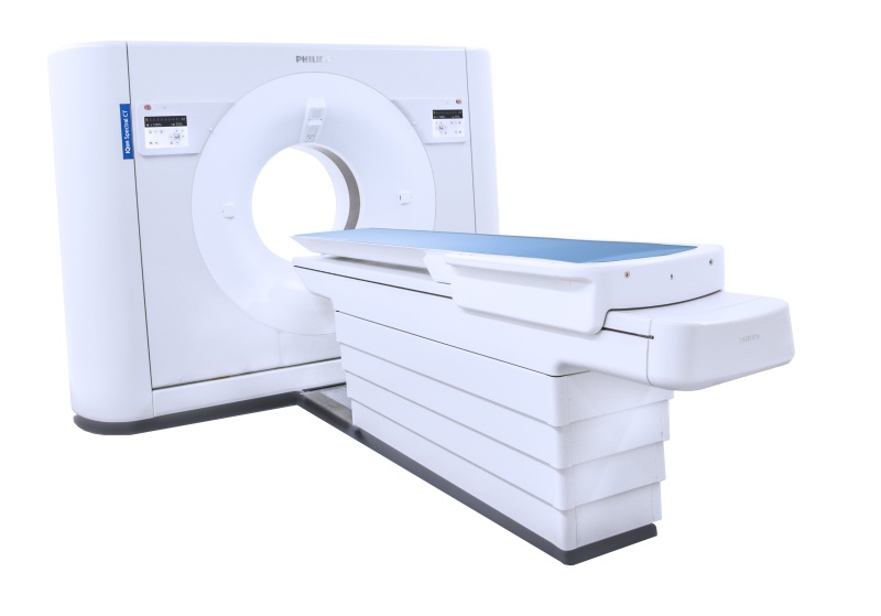
Philips IQon Spectral CT
“As price goes down, spectral will become new normal,” Boussebaa said.
Siemens Healthineers
It’s been a busy year for the Siemens Healthineers CT segment. In April 2018, the company received FDA clearance for the SOMATOM go.All and SOMATOM go.Top systems, the latest addition to its SOMATOM family of CT scanners.
The scanners include a redesigned tablet workflow that allows technologists to spend more time with patients, a development that came after the company engaged with more than 500 customers from around the world in co-creation sessions.
“We believe this is a major enhancement,” said Matthew Dedman, U.S. marketing director for CT for Siemens Healthineers North America.
Along with the tablet workflow comes intelligent automation, including the ability to set up the scan range and the reconstruction field of view automatically, for greater standardization and consistency of results, Dedman said.
The scanners also come with a larger 75-kilowatt generator, for busier and more complex environments.
“We enhanced the horsepower of those systems to enable more advanced exams, such as emergency department patients and cardiac,” Dedman said.

Siemens SOMATOM Edge Plus CT system
“The industry as a whole has done a good job in investing in iterative reconstruction,” Dedman said. “Additionally, we have invested heavily into hardware-based dose reduction technologies as well. If we can optimize our hardware, so that prior to any iterative reconstruction we are delivering the lowest dose, high-quality image, that means we’re less reliant on IR algorithms and can maintain a more natural image impression for the radiologist.”
In April, the company also received FDA clearance for its SOMATOM Edge Plus and SOMATOM Force CT systems. Both scanners feature Fully Assisting Scanner Technologies (FAST) Integrated Workflow with what the company calls the FAST 3D Camera, a patient positioning system that uses artificial intelligence.
Historically, patient positioning has been a manual task, with the height of the technologist impacting the patient position, Dedman explained.
“A six-four tech versus a five-six tech will have a different visual perception of what the ISO center is,” Dedman said.
The FAST 3D camera, mounted above the table, uses an AI algorithm to do correct positioning for the exam, reducing variability among exams.
“When we looked at areas to develop new technologies, we’re always looking for ways to improve standardization and increase consistency for our customers,” Dedman said.
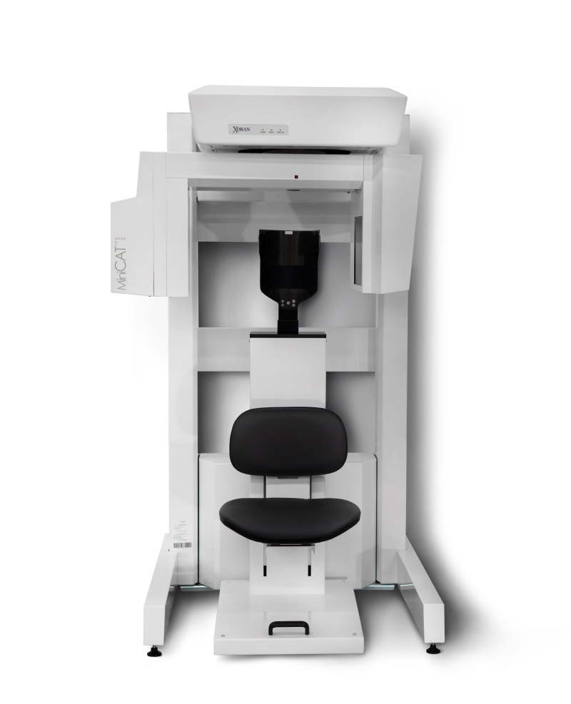
Xoran MiniCAT 2020
Xoran specializes in point-of-care CT scanners that are utilized in doctor's offices as well as the operating room. The company’s flagship product is the MiniCAT, which is primarily used in ENT and allergy offices for CT scans of the ears and sinuses.
In April 2018, the company released the MiniCAT 2020, a scanner that is further optimized for CT imaging within the workflow of the ENT clinic.
The scanner can differentiate between bone, liquid and air, giving clinicians the ability to detect disease, infection, obstruction and other ailments, said Russell Jahnke, director of product management for Xoran Technologies.
“We really looked at what patients are expecting,” Jahnke said. “When they go see their ENT, they really want to see things taken care of in as few visits as possible. The point-of-care imaging the MiniCAT offers in the ENT office is exactly that.”
In the middle of last year, the company introduced the xCAT, a portable head CT scanner for use in the OR, allowing surgeons access to CT imaging during surgery. This year, Xoran also released the xCAT IQ, a portable intraoperative CT designed for neurosurgeons to use in both the OR and the ICU.
“If a neurosurgery patient is in the ICU, the current standard-of-care is to pack them up and take them to the CT suite,” Jahnke said. “With the xCAT IQ, you can bring the device to the bedside.”
Earlier in 2018, Xoran debuted what it calls “Ace Mode” on its MiniCAT IQ sinus and temporal bone scanner used in the doctor's office. When MiniCAT is in ACE Mode, it uses patient-specific data obtained during the pre-scan setup to automatically present a patient profile that helps the clinician select the just-right patient CT scan.

Zetta Medical Z-DOSERP
In July 2017, Zetta Medical Technologies, a third-party service provider that also develops CT software, released Z-DOSE RP, a dose reporting platform. The software connects with the ACR dose registry and also allows facilities to track dose data by study type over multiple scanners and locations.
“A lot of the legacy systems don’t have those capabilities,” said Mike Ghazal, the chief executive officer of Zetta Medical Technologies. “Even the new scanners will generate a structured report, but don’t generate a dose report for the director of radiology to view and track data. Ours is more of a dashboard comparison to analyze dose data over time using multiple criteria. In addition to CTDi and DLP values, it also includes comparisons by exam type, scanner type, referring physician, etc.”
The product is designed for large university hospitals and imaging centers, and is designed to work with the scanners from the major manufacturers, including Canon, GE, Philips and Siemens.
“Once they see the reports, they don’t want to go back to not using it,” Ghazal said. “They like the ability to see all this data and have it at their fingertips.”
The company has designed custom templates for its customers to allow them to view more data to their own specifications.
“Every place has its own set of protocols,” Ghazal said.