
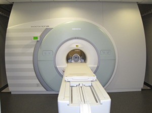
Kamil Ugurbil, director of the University
of Minnesota CMRR, stands beside the
10.5 Tesla MR from Siemens Healthineers.
of Minnesota CMRR, stands beside the
10.5 Tesla MR from Siemens Healthineers.
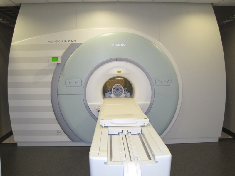
Pushing boundaries of medical imaging with 10.5 Tesla MR
September 10, 2018
by Gus Iversen, Editor in Chief
In February, after many years of planning, the Siemens Healthineers 10.5 Tesla MR at the University of Minnesota’s Center for Magnetic Resonance Research performed a first-ever 10.5 Tesla full scan of the human body.
The hope is that this machine, with its 110-ton magnet, will provide fresh insight into brain function as well as a range of illnesses, such as heart disease, diabetes, and cancer.
HealthCare Business News contacted the team at UMN and asked them a few questions about the project. The responses, which were provided by email, come from Kâmil Uğurbil, director of the CMRR, and associate professors Greg Metzger and Gregor Adriany.
HCB News: How did UMN come to acquire a 10.5 Tesla MR system?
The 10.5 Tesla project at the Center for Magnetic Resonance Research (CMRR) was funded by an $8 million High End Instrumentation grant from the NIH, with additional support from the University of Minnesota toward acquisition of the magnet and construction of the space for the system.
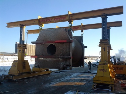
The grant aimed to push the boundaries of MR technology further by establishing a 10.5 Tesla MR imaging and spectroscopy instrument with a sufficiently large bore size (83 cm clear bore) to perform studies on the human body. This instrument was to be first of its kind in the world and the highest field available for research in the human brain as well as the human torso and extremities.
HCB News: What are the shielding specifications like for a magnet that strong? Did the very high strength require special considerations?
There are two ways to reduce the fringe field associated with such an ultrahigh-field magnet to acceptable levels (~5 gauss), either use a large magnet to counteract the magnetic field (actively shielded magnet) or surround the magnet with a massive iron housing (passively shielded magnet). For the CMRR’s 10.5 T the only option was to use a passive shield using 600 tons of iron.
Building an active shield would have made the magnet too large and heavy as it is integrated into the same cryostat (super cooled part of the magnet) as the windings used to generate the desired 10.5 T used for imaging.
The iron shielding reduces the magnetic field from inside the magnet of 10.5 T to ~5 gauss at the perimeter (~7 m from the enclosure). In the absence of the passive shield, the 5 gauss line would have been much further away; much too far to site at the CMRR considering the other equipment in the facility (other high-field systems, a levitation magnet, optical imaging labs, cyclotron. etc.).
HCB News: What kind of research did you start off doing with the system prior to scanning the first human patient?
Extensive preclinical imaging was performed to investigate the vestibular, cognitive and physiologic effects of exposure to ultrahigh magnetic fields. The data supported an Investigational Device Exemption (IDE) application submitted to the FDA in order to justify an initial safety study to be performed on this one-of-a-kind system.
The IDE and subsequent safety study were required as magnets with field strengths greater than 8 Tesla are no longer considered non-significant risk per FDA guidelines, “Criteria for Significant Risk Investigations of Magnetic Resonance Diagnostic Devices”. Only after the safety study is completed and results are positively reviewed by the FDA will broader clearance be given for more specific studies to investigate the system’s capabilities and pursue answers to biomedical questions.
HCB News: How was the first human patient chosen?
 The first human imaging studies were performed on healthy controls that passed the strict screening procedure of the ongoing FDA and local IRB (Institutional Review Board) approved safety study. In this initial cohort of subjects, physiological parameters need to be within normal ranges and a health questionnaire is administered and reviewed by a physician to rule out individuals with pre-existing conditions that meet specific exclusion criteria. In addition, subjects need to be free of any metallic implants while meeting both age and weight requirements.
HCB News: What were the first imaging studies performed and how would you describe the image quality compared to a conventional 3 T MR scan?
To date, the studies that have been performed have been in the human torso. This general anatomical region has been the focus, due to the ability to more rapidly validate radiofrequency (RF) coils, which are heavily loaded by the body. Our body array coils consist of elements that both transmit and receive RF energy from the body (i.e., transceivers). Each element is pressed against the body, facilitated by a flexible housing. For head imaging, a similar flexible housing is not feasible, therefore the elements can have varying distance from the head, making the validation of head RF coils more time-consuming.
The imaging studies performed to date have included the prostate and bilateral hip imaging, target anatomies for which we have experience at 7 T. While very little time is available for imaging, and protocol optimization is only possible in phantoms, we have been able to obtain high quality images of both targets.
 The RF coil and RF shimming methods used to manage the transmit fields worked especially well and allowed for us to obtain high-quality gradient echo images in all cases while fat suppression was feasible within nearly the entire field of view due to the increased chemical shift dispersion at 10.5T, despite the increased B0 inhomogeneities. For prostate imaging, we were able to obtain multi-slice T2-weighted images using a fast spin echo imaging approach. For the musculoskeletal applications, we obtained high resolution 0.7 mm isotropic versions of standard 3D acquisitions, although the optimization of parameters to obtain the desired contrasts require further investigation.
HCB News: Did special RF coils and gradients need to be developed for the system? Are those components continuing to be developed in order to maximize the scanner's capabilities?
For the gradients we are utilizing a high-performance but standard clinical body gradient (Siemens, SC 72). There are also higher-performance commercial head gradients available (AC84) for the system, but in order to further increase the scanner’s maximal spatial and temporal resolution beyond the capabilities of the AC84 head gradient we are developing a novel head gradient insert in collaboration with Stanford University as part of an NIH research grant.
For all our imaging applications we develop special UHF RF coils operating at 447 MHz for protons at 10.5 T. Regarding RF coils, the path to maximize the scanner capabilities will be by extending the number of receiver channels to 128 and the number of independent transmitters to 32.
HCB News: Are there specific biomedical applications or indications/diseases for which you can see 10.5 T MR offering clear benefits and perhaps justifying the arrival of such a powerful magnet on the clinical, commercial level?
There are many potential advantages accompanying the increase in static field strength to 10.5 T that justify further developing the technology and assessing its safety for human investigations. First, it has been shown that sensitivity, as measured by the signal-to-noise ratio (SNR), scales supralinearly with the field strength. While acquisition and anatomy-specific relaxation effects need to be accounted for, this underlying increase in SNR plays an important role in the overall benefits expected from 10.5 T compared to lower field strengths.
Beyond the increase in SNR, other advantages include increasing susceptibility-based contrast for improved anatomic and functional imaging, increased chemical shift dispersion for improved spectroscopic quantification, shifting exchange to faster regimes for improved chemical exchange saturation transfer (CEST) studies, and increased longitudinal relation times for improved non-contrast-enhanced arterial spin-labeled (ASL) perfusion and in-flow angiography.
 One of the initial motivations pushing the development of the 10.5 T scanner was the drive to image functional structures in the brain with increasing spatial resolution and fidelity. Accomplishing this with functional magnetic resonance imaging (fMRI) would provide unprecedented access to small (submillimeter) organizations critical in function of the brain, such as cortical layers and so called cortical columns that represent elementary computational units. With significant developments in auxiliary technologies that exploit the 10.5 T field strength, which we are currently undertaking, we expect to obtain functional images that span elementary computational units to whole brain coverage.
HCB News: What plans do you have for the system in the coming months or years?
Our plans are to complete the safety study and receive approval to perform more extensive studies exploring the true potential of the applications we just discussed.
HCB News: Looking back from today to the beginning of the 10.5 Tesla MR project, what has been the most rewarding or surprising aspect of this endeavor?
The most rewarding aspect has been that 10.5 T appears to be poised to deliver our expected gains in sensitivity (i.e., SNR), and that many of the technologies we have been developing at 7T, with some modification are able to solve the difficulties encountered at 10.5 T.
 As the research site that launched the 7T effort we pursued many of the fundamental studies aimed at understanding the RF interactions with human bodies at high frequencies, which are proving generalizable to 10.5T and likely even higher magnetic fields. With the promise of this system close to being realized, the most surprising aspect is the fact that it took so long to achieve from conception to first human studies (about a decade under development).
The hope is that this machine, with its 110-ton magnet, will provide fresh insight into brain function as well as a range of illnesses, such as heart disease, diabetes, and cancer.
HealthCare Business News contacted the team at UMN and asked them a few questions about the project. The responses, which were provided by email, come from Kâmil Uğurbil, director of the CMRR, and associate professors Greg Metzger and Gregor Adriany.
HCB News: How did UMN come to acquire a 10.5 Tesla MR system?
The 10.5 Tesla project at the Center for Magnetic Resonance Research (CMRR) was funded by an $8 million High End Instrumentation grant from the NIH, with additional support from the University of Minnesota toward acquisition of the magnet and construction of the space for the system.
The grant aimed to push the boundaries of MR technology further by establishing a 10.5 Tesla MR imaging and spectroscopy instrument with a sufficiently large bore size (83 cm clear bore) to perform studies on the human body. This instrument was to be first of its kind in the world and the highest field available for research in the human brain as well as the human torso and extremities.
HCB News: What are the shielding specifications like for a magnet that strong? Did the very high strength require special considerations?
There are two ways to reduce the fringe field associated with such an ultrahigh-field magnet to acceptable levels (~5 gauss), either use a large magnet to counteract the magnetic field (actively shielded magnet) or surround the magnet with a massive iron housing (passively shielded magnet). For the CMRR’s 10.5 T the only option was to use a passive shield using 600 tons of iron.
Building an active shield would have made the magnet too large and heavy as it is integrated into the same cryostat (super cooled part of the magnet) as the windings used to generate the desired 10.5 T used for imaging.
The iron shielding reduces the magnetic field from inside the magnet of 10.5 T to ~5 gauss at the perimeter (~7 m from the enclosure). In the absence of the passive shield, the 5 gauss line would have been much further away; much too far to site at the CMRR considering the other equipment in the facility (other high-field systems, a levitation magnet, optical imaging labs, cyclotron. etc.).
HCB News: What kind of research did you start off doing with the system prior to scanning the first human patient?
Extensive preclinical imaging was performed to investigate the vestibular, cognitive and physiologic effects of exposure to ultrahigh magnetic fields. The data supported an Investigational Device Exemption (IDE) application submitted to the FDA in order to justify an initial safety study to be performed on this one-of-a-kind system.
The IDE and subsequent safety study were required as magnets with field strengths greater than 8 Tesla are no longer considered non-significant risk per FDA guidelines, “Criteria for Significant Risk Investigations of Magnetic Resonance Diagnostic Devices”. Only after the safety study is completed and results are positively reviewed by the FDA will broader clearance be given for more specific studies to investigate the system’s capabilities and pursue answers to biomedical questions.
HCB News: How was the first human patient chosen?

A greater signal-to-noise (SNR) ratio is a key benefit high
field strength magnets may have over conventional MR systems.
field strength magnets may have over conventional MR systems.
HCB News: What were the first imaging studies performed and how would you describe the image quality compared to a conventional 3 T MR scan?
To date, the studies that have been performed have been in the human torso. This general anatomical region has been the focus, due to the ability to more rapidly validate radiofrequency (RF) coils, which are heavily loaded by the body. Our body array coils consist of elements that both transmit and receive RF energy from the body (i.e., transceivers). Each element is pressed against the body, facilitated by a flexible housing. For head imaging, a similar flexible housing is not feasible, therefore the elements can have varying distance from the head, making the validation of head RF coils more time-consuming.
The imaging studies performed to date have included the prostate and bilateral hip imaging, target anatomies for which we have experience at 7 T. While very little time is available for imaging, and protocol optimization is only possible in phantoms, we have been able to obtain high quality images of both targets.
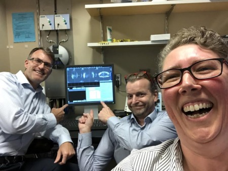
Imaging with the 10.5 T system was an achievement
nearly a decade in the making.
nearly a decade in the making.
HCB News: Did special RF coils and gradients need to be developed for the system? Are those components continuing to be developed in order to maximize the scanner's capabilities?
For the gradients we are utilizing a high-performance but standard clinical body gradient (Siemens, SC 72). There are also higher-performance commercial head gradients available (AC84) for the system, but in order to further increase the scanner’s maximal spatial and temporal resolution beyond the capabilities of the AC84 head gradient we are developing a novel head gradient insert in collaboration with Stanford University as part of an NIH research grant.
For all our imaging applications we develop special UHF RF coils operating at 447 MHz for protons at 10.5 T. Regarding RF coils, the path to maximize the scanner capabilities will be by extending the number of receiver channels to 128 and the number of independent transmitters to 32.
HCB News: Are there specific biomedical applications or indications/diseases for which you can see 10.5 T MR offering clear benefits and perhaps justifying the arrival of such a powerful magnet on the clinical, commercial level?
There are many potential advantages accompanying the increase in static field strength to 10.5 T that justify further developing the technology and assessing its safety for human investigations. First, it has been shown that sensitivity, as measured by the signal-to-noise ratio (SNR), scales supralinearly with the field strength. While acquisition and anatomy-specific relaxation effects need to be accounted for, this underlying increase in SNR plays an important role in the overall benefits expected from 10.5 T compared to lower field strengths.
Beyond the increase in SNR, other advantages include increasing susceptibility-based contrast for improved anatomic and functional imaging, increased chemical shift dispersion for improved spectroscopic quantification, shifting exchange to faster regimes for improved chemical exchange saturation transfer (CEST) studies, and increased longitudinal relation times for improved non-contrast-enhanced arterial spin-labeled (ASL) perfusion and in-flow angiography.
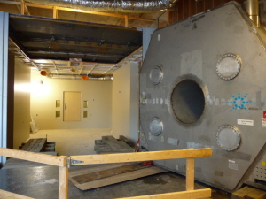
The passive shielding for the 10.5 Tesla scanner
required 600 tons of iron.
required 600 tons of iron.
HCB News: What plans do you have for the system in the coming months or years?
Our plans are to complete the safety study and receive approval to perform more extensive studies exploring the true potential of the applications we just discussed.
HCB News: Looking back from today to the beginning of the 10.5 Tesla MR project, what has been the most rewarding or surprising aspect of this endeavor?
The most rewarding aspect has been that 10.5 T appears to be poised to deliver our expected gains in sensitivity (i.e., SNR), and that many of the technologies we have been developing at 7T, with some modification are able to solve the difficulties encountered at 10.5 T.

Funded by an $8 million grant from the NIH, the researchers at the UMN Center
for Magnetic Resonance Research are now conducting 10.5 T studies on the human body
for Magnetic Resonance Research are now conducting 10.5 T studies on the human body